What is it?
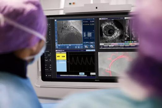
Intravascular Ultrasound Study is an invasive diagnostic procedure that involves creating real-time images of the coronary arteries as well as its inside, using ultrasound frequency. This gives the doctor a detailed visual image of the arteries, which makes it easier to identify and diagnose blockages in the arteries or narrowing of the walls. It is also often used to check the efficiency of angioplasty or arthrectomy procedure done, as well as in the placement procedure of stents. In rare cases of a re-stenosis (reoccurrence of buildup in arteries), Intravascular Ultrasound can prove beneficial to quickly identify it, making it easier to propose a treatment immediately.
How is it done?
Intravascular Ultrasound Study is an invasive procedure, and hence involves the use of anesthetic being administered, before prepping the required site for insertion of the apparatus. Once the patient is under sedation, a catheter is slowly inserted through the targeted region and guided into the arteries, to locate the problem area. The catheter is fitter with a small camera-like device which helps create detailed images of the arteries, through ultrasound waves emitted from it. Once the necessary images are obtained, the apparatus is safely removed and the patient is bandaged up.
Patients will need to arrange for a ride back, post-procedure, as well as follow the steps instructed by the doctor. These could include getting adequate rest, staying hydrated, eating healthy, etc.
Why is it done?
Intravascular Ultrasound Study can help with various functioning, including:
- Identifying and diagnosis narrowing or thickening of artery walls
- Locating blockages in the arteries
- Creating images of the arteries before, during and/or after an angioplasty or arthrectomy
- Assisting stent placement procedures
- Identifying reoccurring buildup and blockage in the arteries


