What is it?
An echocardiogram is a diagnostic procedure that involves developing a graphic outline of the heart’s activity using ultrasound frequency. The images created by the machine can help doctors understand the heart’s pumping action and look out for any discrepancies in the heart’s functioning. An echocardiogram can be done using various techniques, including:
- Two-dimensional ultrasound
- Three-dimensional ultrasound
- Doppler ultrasound
- Color Doppler ultrasound
- Strain imaging
- Contrast imaging
Out of these techniques, the most commonly used one, is the 2D ultrasound echocardiogram. This helps create 2D images of the heart that appear as thin slices on the computer screen, depicting the heart’s pumping.
There are various types of echocardiograms, namely:
- Transthoracic echocardiogram
- Transesophageal echocardiogram
- Exercise stress echocardiogram
An echocardiogram can often take anywhere from 40 to 60 minutes, depending on the type of procedure done as well as the extent of readings taken. A transesophageal echocardiogram, for instance, can take up to 90 minutes.
How is it done?
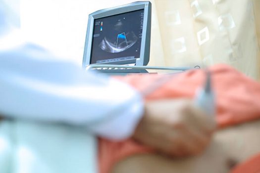
During an echocardiogram, the doctor or technician makes use of a hand-held device, known as a transducer, to create the 2D images of the heart. The device, emitting ultrasound frequency, is placed near the outside of your heart, sending the frequency waves through. These waves, upon hitting the heart, bounce back like echoes, which is then received and translated on the computer screen as 2D images.
While preparing for an echocardiogram, you will be briefed on what you can or cannot do. Although there are little to no steps needed to be taken to prepare for the procedure, it is beneficial to understand the procedure and expect the following:
- You will be asked to remove clothing from the upper half of your body, and wear a hospital gown. This helps make it easier for the doctor to reach the transducer towards the chest.
- You may be fitted with ECG electrodes as well, giving the doctor an ECG reading along with the echocardiogram images.
- The transducer will be lubricated with a gel to make it easier to glide on the skin. This gel is harmless to the skin and you need not be worried.
Once the procedure is done, the results will be analyzed by the doctor and conveyed to you during the follow-up sessions.
Why is it done?
An echocardiogram can help doctors identify and diagnose various heart-related problems, including:
- Congenital heart disease
- Cardiomyopathy
- Infective endocarditis
- Pericardial disease
- Valve disease
- Aortic aneurysm
- Blood clots
- Cardiac tumors


